


What is Osgood Schlatter’s Disease?
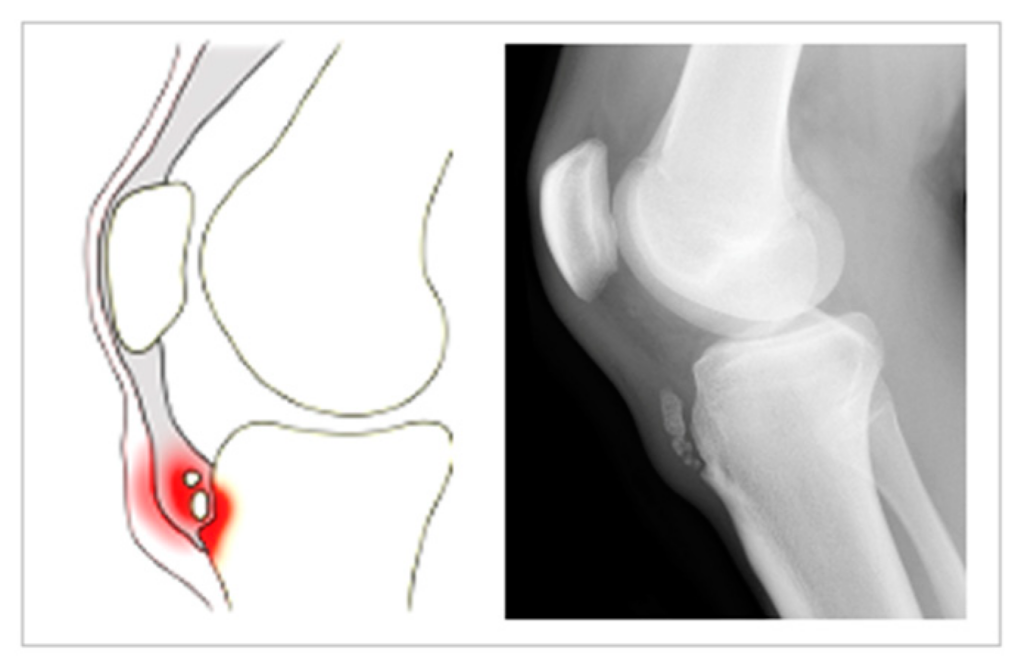 It is an overuse injury at the site of the tibial tuberosity, on the tibial bone, where the patellar tendon attaches. It is apophysitis (inflamed bone at the site of the tibial tuberosity growth plate) of tibial tuberosity seen in children.
Though it is relatively uncommon, it is a quite debilitating knee injury that occurs in young active children/adolescents.
Activities that involve repetitive, strong quadriceps contractions, such as jumping, running, volleyball, basketball, soccer, gymnastics, dance, netball, and ice skating are most at risk.
Symptoms of Osgood Schlatter’s Disease
Osgood Schlatter’s disease presents in growing boys and girls as :
- Local pain, swelling, and tenderness over the tibial tuberosity
- Pain is experienced during exercise (e.g. running, jumping) or with direct contact, such as in kneeling.
- Pain in activities like stair climbing, squatting, and kneeling.
- Quadriceps weakness can be present in chronic cases.
- Bilateral symptoms occur in 20-30% of cases.
- The apophysis may be enlarged in later stages, which looks like a lump that is tender in its active phase.
Causes
Strong repetitive quadriceps contractions are thought to cause a traction force on the tibial tuberosity, disrupting the immature bone. There is a higher incidence of inactive children during the adolescent growth spurt.
During the development phase of bone, it goes through different stages as–
- The tibial tuberosity is initially cartilaginous (cartilaginous stage).
- It then enters the apophyseal stage when the secondary ossification center (apophysis) appears.
- The unity of the proximal tibial epiphysis with the tibial apophysis marks the epiphyseal stage and
- Lastly, when the growth plates fuse, the bony stage has been reached.
Children are most susceptible to Osgood-Schlatter’s disease when their bones are in the (2nd) apophyseal stage.
During this stage, the apophysis is unable to withstand high tensile forces. When presented with strong, repetitive muscle contractions, micro-fractures occur in the immature area. The separation results in symptoms typical to Osgood Schlatter’s disease, as well as irregular bone growth that explain an enlarged tibial tuberosity afterward.
Another reported cause for Osgood Schlatter’s disease is mismatched growth of the quadriceps in comparison to the femur. During a growth spurt in a child, the lengthening of the muscle is unable to keep up with the rapid lengthening of the femur bone, resulting in an increased tensile force on the tibial tuberosity.
The most prevalent groups are Boys: ages 11-15 years – Girls: ages 8-13 years.
Diagnosis
Osgood Schlatter’s Disease can normally be diagnosed by careful examination and looking for signs and symptoms related to the condition.
Plain X-rays are commonly taken to rule out other conditions such as a tibial tuberosity fracture, malignancy, or infection.
MRI is not commonly used to confirm the diagnosis.
Treatment of Osgood Schlatter’s Disease
- Physiotherapy
Physiotherapy treatment is beneficial in Osgood Schlatter’s disease. About 90% of patients respond to non-operative physiotherapy treatment, but symptoms may come and go for 12-24 months before complete resolution.
Knee Protection is an important part of treatment. This includes –
- Immediate restriction of high-impact activities such as jumping and running.
- Use of infrapatellar knee strap to restrict force transmission to the site of Osgood Schlatter’s Disease.
- Kinesiology taping provides both pain relief and load reduction at the site of pain and injury.
- Severe cases of Osgood Schlatter’s disease may require crutches.
- Pain and inflammation control
A combination of ice and TENS treatment will help reduce pain and inflammation. It improves the healing rate.
Rest is important in the management of Osgood Schlatter’s disease and the relief of pain. Exercises and sporting activities are stopped during the flare-up stage of the disease.
- Therapeutic Exercises
Stretching and Massage
One of the common reasons for developing Osgood Schlatter’s disease is excessively tight quadriceps muscles, ITB, hamstrings, hip flexors, and calf muscles. The physiotherapist will design stretching exercises for these muscles as well as teach the patient how it can be done at home.
Massage/ foam rollers especially in the early phase when stretches create pain at the Osgood Schlatter’s disease site can be beneficial.
Strengthening
The muscle balance around the knee may not be adequate and causing excess stress at the site of the tibial tuberosity. A physiotherapist will help in designing an appropriate strengthening regime.
Foot and Arch Control & Orthotics
The foot biomechanics or arch control may be inadequate in an individual for the high intensity of the sport.
The physiotherapist can assist in both the assessment and corrective exercises for dynamic foot control.
Prognosis:
It is a self-limiting disease in which complete recovery can be expected with the closure of the tibial growth plate. In the long run patients with an enlarged lump at the site as a result of apophysitis may complain of pain with kneeling activities.
The condition in some cases may linger on for months and doesn’t show good recovery with conservative treatment and may need surgical intervention. Corticosteroid injections cause’ subcutaneous atrophy hence not recommended.