What are the Reasons for Posterior Tibialis Tendonitis/Rupture
The Reasons for Posterior Tibialis Tendonitis/Rupture are as follows:
- Overuse activities: Posterior Tibialis Tendonitis/Rupture is usually an Overuse Injury, which commonly occurs due to repetitive or prolonged activities placing strain on the tibialis posterior tendon. This typically occurs due to excessive walking or running (especially up slopes or on uneven surfaces), jumping, hopping, or change of direction activities.
- Trauma: Patients may develop this condition suddenly due to a forceful contraction of the tibialis posterior muscle often when in a position of stretch. This typically occurs during rapid acceleration while running (particularly when changing direction).
- Degenerative: Sometimes the tendon may have degeneration within it as well and not just inflammation. Degeneration typically occurs in middle-aged and older individuals.
Tibialis posterior
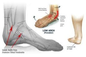
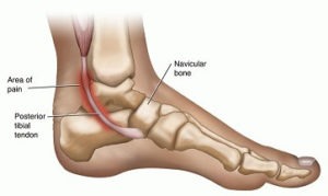
The tibialis posterior muscle originates from the back of the tibia and fibula (lower leg bones); it then runs down along the inside of the lower leg and ankle (behind the medial malleolus) and inserts into various bones in the foot via the tibialis posterior tendon. The tibialis posterior muscle is responsible for rotating the foot and ankle towards the midline of the body (inversion) and pointing the foot and ankle down (plantarflexion). In cases of dysfunction or injury, pain free physiotherapy can help restore mobility, reduce discomfort, and improve overall function.
It is one of the major supportive structures of the foot and active mainly during weight-bearing activities. The tendon helps to keep the arch of the foot in its normal position. When there is insufficiency or rupture of the tendon, the arch begins to sag and a flat foot deformity can occur with associated tight Achilles tendon. The posterior tibial tendon rupture occurs distal to the medial malleolus.
This area is hypovascular.
Clinical presentation:
- Painful swelling on the posteromedial aspect of the ankle.
- Unable to perform single-leg toe raise.
- Too many toe signs.
- Flat foot deformity.
- Fixed deformity of the hindfoot.
- Rupture of the posterior tibial tendon could be missed.
Stages of Posterior Tibialis Tendonitis/Rupture :
There are four stages of posterior tibial tendon insufficiency.
- The first stage refers to inflammation of the tendon in which no deformity has occurred. This is not common.
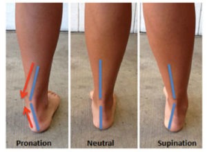
- Usually, patients present with stage II in which flexible deformities occur. The arch begins to collapse and there is increased hindfoot valgus. In stage II, there is no significant forefoot abduction. However, in stage II b significant forefoot abduction is seen.
- In stage III, the deformity has progressed long enough to where arthritis develops in the hindfoot joints leading to a stiff deformity.
- In stage IV, the involvement of the tibiotalar joint in addition to the hindfoot malalignment is seen. This most advanced stage is comprised of a hindfoot valgus deformity, resulting from degeneration of the posterior tibial tendon, with associated valgus tilting of the talus within the mortise.
Treatment Of Posterior Tibialis Tendonitis/Rupture :
- Conservative treatment
As with most cases of soft tissue injuries the initial treatment is – Rest, Ice, and Protection.
- Rest: In the early phase the best is to avoid all activities that induce pain.
- Ice is a simple and effective modality to reduce pain and swelling. Apply ice for 10-15 minutes every 2 to 4 hours during the initial phase.
- Protection: the Physiotherapist may normally apply supportive taping or ankle binder to help relieve pain and commence the realignment phase. Taping is normally immediately effective in providing pain relief.
- Anti-inflammatory medication and creams may help reduce pain and swelling.
- A cortisone injection is usually not appropriate for this condition, since the tendon is more likely to rupture following injection. Alternatively, iontophoresis with cortisone ointments can be recommended. Iontophoresis is a treatment that uses electric current to deliver medically active ions through the skin.
During the initial treatment, Physiotherapy aims at reducing pain and swelling. Apart from rest, ice, and protection physiotherapist can use therapeutic modalities like ultrasound therapy, iontophoresis, laser therapy TENS in any possible combination to achieve this. The Physiotherapist can use myofascial release at the bottom of the foot, calf, and muscles around the shin. This is useful in relieving the discomfort through a proper excursion of the muscles. These treatment options are particularly helpful if patients present with tendonitis without any bony changes like an arch drop. In advance stages of tibialis posterior insufficiencies, physiotherapist needs to address the problems of why the posterior tibial tendon is being stressed will provide more long-term relief to the injury and help to avoid an irritated tendon from developing into a chronic problem or rupture.
Physiotherapy exercises to strengthen the small muscles of the foot and posterior tibialis muscle. As mentioned above, these muscles work to lift and support the arch of the foot, along with the muscles of the hip, knee, and ankle which control the alignment down the lower leg chain and also assist in lifting the arch of the foot. Biomechanical correction of foot and ankle along with strong pelvic stabilization is needed for proper weight transfer and absorption of ground reaction forces. This will minimize the stresses on the posterior tibial muscle.
Stretching exercises for the calf, hamstring, and intrinsic muscles of the foot which when tight can force the foot out of alignment. When performing all stretches it is important to maintain proper alignment of the foot and arch so as not to stress the already irritated posterior tibial tendon.
The posterior tibial tendon problems develop over time and it is common for patients to overlook the problem unless pain appears. Re-aligning the arch of the foot to the proper position is crucial to relieve the pain in the posterior tibial tendon as well as stop the progression of the arch deformation. Proprioceptive exercises are taught to the patient to increase the sense of joint positions of lower limb joints. Initially, these are practiced in sitting positions and gradually in more challenging positions.
The physiotherapist should advise the patient proper footwear with arch support orthotic aid to take off a load of the posterior tibial tendon. Often taping the foot can be performed by the physiotherapist and can be taught to the patient as well, to be applied when the arch support is not on. Taping may be enough in mild cases of posterior tibial tendon pain, as long as the patient learns to control the position of the foot and maintain this position during high-level activities. In most cases, shoe inserts, even pre-fabricated ones, will significantly improve the symptoms of a posterior tibial tendon problem and will be recommended to both relieve symptoms and avoid any progression of the injury. Custom-fit orthotics are recommended for any individuals who have a significant flattened arch, or for whom prefabricated ones do not relieve their symptoms.
Excess weight can be a cause of posterior tibial tendon problems. Losing excess weight can greatly improve the pain felt in the foot and make it easier to maintain normal foot alignment during everyday activities. Often it is difficult to exercise to lose weight with the painful feet, however, there are several safe activity options, such as stationary cycling or swimming.
Not all conditions of posterior tibial tendonitis/ insufficiencies are managed conservatively and may need an Orthopaedician opinion for more suitable options of surgical remedies.
- SURGERY:
If all else fails to resolve your condition, surgery may be required.
Surgical debridement (thickened and inflamed tissue removal arthroscopically), tendon repair for degenerated tibialis posterior, tendon transfer (flexor digitorum longus tendon is transferred to perform the action of tibialis posterior in cases of rupture), and arthrodesis of talonavicular, talocalcaneal, and calcaneocuboid joint (triple arthrodesis) in severe cases of flat feet deformity (stage IV) are few surgical resorts to resolve posterior tibialis injuries.
- Post-surgical rehabilitation:
Post-surgical rehabilitation may take 4-6 weeks time to get back to normal depending upon the surgical method implemented. In simple debridement and tendon repair cases it may occur in a relatively lesser time period and treatment will remain similar to conservative treatment.
Among the tendon transfer case reeducation of the transferred tendon is the most essential part which needs to be emphasized. The rest of the treatment remains more or less the same as conservative treatment which aims at reduction of pain post-operative with therapeutic modalities, weight reduction, proprioceptive exercises, strengthening the muscles around the ankle, knee, hip, and pelvic stabilization exercises, biomechanical correction of ankle and foot and appropriate orthotic aid for arch support to reduce the tension on the newly transferred tendon.
Rehabilitation of arthrodesis cases needs a relatively long time period of 12-15 weeks. The orthotic aid is gradually weaned off after 3 months period as the surgeon feels the arthrosed joints are capable of bearing the weight. The patient is on non-weight bearing till 6-8 weeks and uses a crutch or walker during this period. Partial weight-bearing is started after 8 weeks with orthotic aid on. Active range of motion exercises of hip, knee, and ankle are started post removal of stitches (10-14 days). Gradual progression to core strengthening and resisted range of motion exercises for lower limb muscles are started as the pain subsides and the patient’s condition improves. The physiotherapist should mobilize the unfused joints of the foot during non-weight bearing. The patient can be made to do strengthening exercises in a pool and gradually learn to bear weight on the affected side in the pool.
As full weight-bearing starts patient is progressed to weight-bearing exercises and Proprioception exercises. The patient can be made to do stationary bicycle. The rest of the exercises is continued as before to achieve need normal status of strength and range of the rest of the joints of the limb.

