Medial Collateral Ligament
The ligament on the medial (inner) side of the knee connecting the femoral condyle and the tibial condyle is called the medial collateral ligament (MCL). It is one of four major ligaments that stabilize the knee joint. It is a flat band of tough connective tissue composed of long, wiry collagen fibers.
The function of MCL is to resist valgus force, which occurs if the tibia/foot is forced outwards in relation to the knee.
Causes of Medial Collateral Ligament Injury
The MCL injury occurs when the (valgus) force is too great for the ligament to resist and the ligament is overstretched. This can occur through-
- A sharp change in direction,
- Twisting the knee whilst the foot is fixed,
- Landing wrong from a jump, or
- A blunt force hit the knee, such as in a football tackle.
The incident IS usually sudden and occurs at high speed. Muscular weakness or incoordination predisposes the ligament to sprains or tear.
Symptoms of Medial Collateral Ligament Injury
Grades of ligament injury
The severity and symptoms of a knee ligament sprain depend on the degree of stretching or tearing of the knee ligament. An audible snap or tearing sound at the time of ligament injury might be the patient’s complaint.

- In a grade I sprain, the ligament is mainly stretched with a minimal tear. There is a little pain, mild swelling, and slight discomfort in weight-bearing activities. A mild ligament sprain can increase the risk of a repeat injury.
- In a grade II sprain, there is a moderate tear (50-70% fibers torn). Severe pain in weight-bearing activities, Swelling, and bruising can be seen around the joint. The feeling of instability i.e. knee giving way medially may or may not be the complaint of the patient
- In grade III sprain, there is a complete tear of the ligament. Swelling and under skin bleed can be seen at times. As a result, the joint is unstable and unable to bear weight. Often there is no pain following a grade 3 tear as all of the pain fibers are torn at the time of injury (pain may be due to injuries to other structures). With these more severe tears, other structures are at risk of injury including the meniscus and/or ACL.

Diagnosis
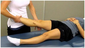
On examination, there may be tenderness over the ligament site, possible swelling, and pain with stress tests.

MRI may be used to confirm the diagnosis and see if any other surrounding structures are not involved.
Treatment of Medial Collateral Ligament Injury
Treatment of an MCL injury depends on the severity and whether there are other concurrent injuries.
- Grade I sprains may heal by themselves in a few week’s time. It may take 6-8 weeks to develop maximum strength in the ligaments (time for collagen fibers to mature). Initial treatment as always in sports injury will be relative rest, icing, compression/ supporting the joint, protection (avoid painful weight-bearing activities). Some NSAIDs can be prescribed by a physician to reduce pain.
Physiotherapy is recommended to increase the healing process. This comprises electrical modalities, soft tissue techniques, strengthening exercises to guide the direction that the ligament fibers heal. This helps to prevent a future tear.
- With a grade II sprain, weight-bearing braces/ supportive taping is a must in early treatment days to relieve pain and avoid stretching of the healing ligament. After a grade II injury, usually returning to activity is possible only once the joint is stable and there is no longer pain. This usually takes six weeks.
Physiotherapy is recommended to hasten the healing process. A physiotherapist uses some modalities, soft tissue techniques, strengthening exercises, and later on, focus on complete rehabilitation so to avoid any factor that may contribute to reoccurrence.
- With a grade III injury, the patient usually wears a hinged knee brace to protect the injury from weight-bearing stresses. The aim is to allow ligament healing and gradually return to normal activities. It may take 3-4 months to completely return to sporting activity. This is again only possible with intensive post-operative physiotherapy rehabilitation.
The aims of physiotherapy treatment are:
- Reduce pain and inflammation.
- Normalize joint range of motion.
- Strengthen the knee muscles.
- Strengthen lower limb muscles
- Improve patellofemoral (kneecap) alignment
- Normalize muscle lengths/ stretches
- Improve proprioception and balance
- Improve functionality e.g. walking, running, squatting, hopping, and landing.
- Guide return to sports activities and exercises
- Minimize re-injury.
However, we strongly suggest that one should discuss his/her knee injury after a thorough examination with a physiotherapist or knee surgeon.
Surgery
Most MCL injuries resolve well with conservative management, however, in grade III injury and if other structures are also involved surgery is considered.
There are few risk factors with every surgery and the ones associated with this include infection, persistent instability and pain, stiffness. However, 90% of patients have no complications post-surgery.
Post-Surgical Rehabilitation
For a successful and quick recovery from knee surgery, rehabilitation is one of the most important aspects.
The physiotherapist focuses on restoring full knee range of motion, strength, power and endurance, balance and proprioception, and agility training that is individualized to specific sports and functional needs.
Prevention
A knee strengthening, agility, and proprioceptive training program is the best way to reduce the chance of a knee ligament sprain. Premature return to high-risk activities such as sport may increase the chance of reoccurrence.


